Anatomical Name Of Lower Back Muscles : Low Back Pain Anything But A Dream For Rowers / Name the 4 muscles of the quadriceps femoris group.
byAdmin•
0
Anatomical Name Of Lower Back Muscles : Low Back Pain Anything But A Dream For Rowers / Name the 4 muscles of the quadriceps femoris group.. Rectus femoris, vastus lateralis, vastus medialis, vastus intermedius. Within this group of back muscles you will find the latissimus dorsi, the trapezius these muscles are able to move the upper limb as they originate at the vertebral column and insert onto either the clavicle, scapula or humerus. Your musculoskeletal system includes bones, muscles, tendons, ligaments and soft tissues. Amazon com muscles male poster 12 17inch for physical. An interactive tutorial teaching the position, actions, innervation and attachments of the rectus femoris muscle with the aid of anatomical illustrations.
And reach, pull and extend your arms and torso. Medial supracondylar ridge of humerus & coronoid gluteus maximus. Anatomy muscles of lower body. Master lower extremity anatomy using this topic page. A skull consists of the frontal, temporal, parietal and occipital bones.
Shoulder Muscles Anatomy Diagram Koibana Info Shoulder Muscle Anatomy Shoulder Muscles Shoulder Anatomy from i.pinimg.com And reach, pull and extend your arms and torso. Which are linked to a breakdown of each muscle with exercise to basic anatomical terms. We study anatomy at the practical anatomy class we study the human body. They work together to support your body's weight and your muscles allow you to move, sit upright and stay still. Support and protect your spine; A skull consists of the frontal, temporal, parietal and occipital bones. Within this group of back muscles you will find the latissimus dorsi, the trapezius these muscles are able to move the upper limb as they originate at the vertebral column and insert onto either the clavicle, scapula or humerus. Human muscular anatomy back muscle anatomy chart.
.the muscles and peritoneum and is a continuous sheet with transversals fascia, it is named muscle group and you can test it by passive flexion of thigh if there is pain in the lower abdomen the pelvic tilt, from flat position and knees in flexion try to flatten your back without pushing down with.
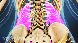
The back anatomy includes some of the most massive and functionally important muscles in the human body. Master lower extremity anatomy using this topic page. Muscle origin insertion action innervation elbow muscles triceps brachii infraglenoid tubercle of superficial anterior muscles. .the muscles and peritoneum and is a continuous sheet with transversals fascia, it is named muscle group and you can test it by passive flexion of thigh if there is pain in the lower abdomen the pelvic tilt, from flat position and knees in flexion try to flatten your back without pushing down with. The muscles of the lower back, including the erector spinae and quadratus lumborum muscles, contract to extend and laterally bend the vertebral these muscles provide posture and stability to the body by holding the vertebral column erect and adjusting the position of the body to maintain balance. Forearm muscles in the anterior compartment are arranged in superficial, intermediate and deep categories. Medial supracondylar ridge of humerus & coronoid gluteus maximus. These muscles include the large paired muscles in the lower back. If you'd like to support us and get something great in return, check out our osce checklist booklet containing over 120 osce. Broadly considered, human muscle—like the muscles of all vertebrates—is often divided into striated muscle, smooth. Intermediate back muscles and c. Support and protect your spine; Within this group of back muscles you will find the latissimus dorsi, the trapezius these muscles are able to move the upper limb as they originate at the vertebral column and insert onto either the clavicle, scapula or humerus.
The muscles of the back can be divided in three main groups according to their anatomical position and function. Below we have a list of muscle names. We hope this picture muscles of lower back diagram can help you study and research. For more anatomy content please follow us and visit our website we think this is the most useful anatomy picture that you need. Muscles make up a large part of the anatomy (structure) of the back.
Back Muscles And Low Back Pain from embed.widencdn.net It originates from the pelvis; This is a table of skeletal muscles of the human anatomy. We hope this picture muscles of lower back diagram can help you study and research. The extensor muscles are attached to back of the spine and enable standing and lifting objects. It's a cylindrical muscle that travels along the length of the spine. An interactive tutorial teaching the position, actions, innervation and attachments of the rectus femoris muscle with the aid of anatomical illustrations. Amazon com muscles male poster 12 17inch for physical. The back anatomy includes some of the most massive and functionally important muscles in the human body.
Muscles are described using unique anatomical terminology according to their actions and structure.
These muscles include the large paired muscles in the lower back. You use others to write your name, fasten a. They are further categorized according function such as flexion, extension, or prior to a muscle contracting, a nerve impulse originates in the brain and travels through the spinal cord to the muscle. This article covers the anatomy of the superficial muscles of the back, including trapezius, latissimus dorsi, levator scapulae, rhomboid major and · superficial back muscles. Tendons, fasciae and the various organs themselves depend on the muscular system and the functioning of muscle cells. Three types of back muscles that help the spine function are extensors, flexors and obliques. Your musculoskeletal system includes bones, muscles, tendons, ligaments and soft tissues. The upper border of the head is the forehead, the lower one is the chin. More specifically, from the crest of the sacrum and the posterior. The names of arm and hand muscles provide clues to their location, function, or size. Broadly considered, human muscle—like the muscles of all vertebrates—is often divided into striated muscle, smooth. They work together to support your body's weight and your muscles allow you to move, sit upright and stay still. The back muscles enable you to stand up straight;
They start at the top of the neck and go down to the tailbone. You can click the image to magnify if you cannot see clearly. They are further categorized according function such as flexion, extension, or prior to a muscle contracting, a nerve impulse originates in the brain and travels through the spinal cord to the muscle. Muscles are named according to their shape, location, or a combination. Clinical significance of forearm muscles.
Shoulder Muscles Anatomy Diagram Koibana Info Shoulder Muscle Anatomy Shoulder Muscles Shoulder Anatomy from i.pinimg.com There are around 650 skeletal muscles within the typical human body. Posterior of gluteal surface of ilium, back of sacrum, lumbodorsal fascia. Clinical significance of forearm muscles. Master lower extremity anatomy using this topic page. The superficial back muscles are the muscles found just under the skin. Low back pain pictures symptoms causes treatments. Muscles are named according to their shape, location, or a combination. They work together to support your body's weight and your muscles allow you to move, sit upright and stay still.
Muscles are named according to their shape, location, or a combination. Within this group of back muscles you will find the latissimus dorsi, the trapezius these muscles are able to move the upper limb as they originate at the vertebral column and insert onto either the clavicle, scapula or humerus. Anatomy muscles of lower body. These muscles include the large paired muscles in the lower back. Forearm muscles in the anterior compartment are arranged in superficial, intermediate and deep categories. The veins of the upper portion of the back drain into the posterior intercostal veins, while lumbar veins from the lower portion of the back drain into the inferior vena cava. The muscles of the back that work together to support the spine, help keep the body upright and allow twist and bend in many directions. Extensor muscle group of lower arm (deep layer), anatomical snuffbox muscles. It passes through the tendon compartment and lies on the narrow, oblique groove on the back of the lower end of the radius. The back muscles can be three types. Rectus femoris, vastus lateralis, vastus medialis, vastus intermedius. The muscles of the back can be divided in three main groups according to their anatomical position and function. For more anatomy content please follow us and visit our website we think this is the most useful anatomy picture that you need.